Pregnancy is not an illness but a most important physiological event in the life of a woman. In most cases,the pregnancy runs smoothly without causing any health problems but you must be under the close supervision of an obstetrician for proper guidance and early identification of any medical problems. You must choose the obstetrician with whom you are comfortable and who is attached to a hospital,which is adequately equipped to provide good quality of newborn care facilities.
You should report to your doctor for first medical check-up as soon as you have missed a period and you feel that you are pregnant. Your doctor will take a detailed history of your past illness and health problems in your family. You must inform your doctor if you have any genital herpes or any sore or ulcer over your genitals at any stage in your life. She will record your date of last menstrual period(LMP) and conduct a thorough examination to assess your nutritional status and any existent health problems.
First examination

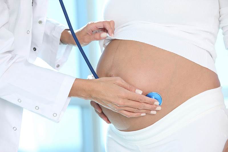
After pregnancy is confirmed, the woman should have a physical examination, preferably between 6 and 8 weeks of pregnancy. At this time, the length of the pregnancy can be estimated and the date of delivery can be predicted as accurately as possible.
Doctors ask about disorders the woman has and has had, drugs she taken, and details about previous pregnancies, including problems that occurred such as diabetes, miscarriages, and birth defects.
The first physical examination during pregnancy is very thorough. It includes the following:
- Blood tests: A sample of blood is taken and analyzed. Analysis includes a complete blood cell count, tests for infectious diseases (such as syphilis, hepatitis, and human immunodeficiency virus [HIV]), and tests for evidence of immunity to rubella and chickenpox (varicella). Blood type, including Rh factor status (positive or negative), is determined.
- Papanicolaou (Pap) test or a variation of it: Samples of tissue from the cervix are taken to check for cancer of the cervix. My doctor have not done this examination at my pregnancy period.
- Test for sexually transmitted diseases: Immediately after the Pap test, another sample of tissue from the cervix is taken to test for sexually transmitted diseases, such as gonorrhea and chlamydial infection.
Other tests may be done, depending on the woman???s situation. If the woman has Rh-negative blood, it is tested for antibodies to the Rh factor . Having Rh antibodies can cause severe problems (even death) for a fetus that has Rh-positive blood. If antibodies in a pregnant woman???s blood are detected early, the doctor can take measures to protect the fetus. All women with Rh 0 -negative blood are given Rh 0 (D) immune globulin, injected into a muscle, at 28 weeks of pregnancy. They are also given an injection after any possible contact between their blood and the fetus’s blood???for example, after an episode of vaginal bleeding or amniocentesis and after delivery.
Follow-up examinations
After the first examination, a pregnant woman should see her doctor every 4 weeks until 28 weeks of pregnancy, then every 2 weeks until 36 weeks, then once a week until delivery. At each examination, the woman???s weight and blood pressure are usually recorded, and the size of the uterus is noted to determine whether the fetus is growing normally. The woman???s ankles are examined for swelling.
Doctors check the heartbeat of the fetus. It can usually be detected at about 10 to 11 weeks with a hand held Doppler ultrasound device. Once a heartbeat has been detected, doctors check it at each visit to determine whether it is normal.
At each visit, urine is tested for sugar. Sugar in the urine may indicate diabetes. If the urine contains sugar, a blood test to check for diabetes is done as soon as possible. Even if the urine does not contain sugar, doctors usually test all women for the type of diabetes that develops during pregnancy (gestational diabetes). This blood test is done at 24 to 28 weeks. It measures the level of sugar (glucose) in the blood 1 hour after women drink a liquid that contains a certain amount of glucose???called a glucose tolerance test. If women have risk factors for gestational diabetes, this test is done early in the pregnancy, preferably before 12 weeks. Risk factors for gestational diabetes include the following:
- Severely overweight (weighing more than 250 pounds)
- Gestational diabetes or a large baby (weighing 10 pounds or more) in a previous pregnancy
- An unexplained miscarriage in a previous pregnancy
- First-degree relatives (such as mothers or sisters) with diabetes
- A history of having sugar in the urine over a long period of time
- Polycystic ovary syndrome with insulin resistance.
If results of the initial test are normal, these at-risk women are retested at 24 to 28 weeks.
At each visit, the urine is also tested for protein. Protein in urine may indicate preeclampsia (a type of high blood pressure that develops during pregnancy???see Preeclampsia and Eclampsia).
If women have a high risk of conceiving a baby with a genetic disorder, prenatal diagnostic testing can be done (see Prenatal Diagnostic Testing).
Ultrasonography
Most doctors believe that ultrasonography (see Ultrasonography), the safest imaging procedure, should be done at least once during a pregnancy to make sure the fetus is normally formed 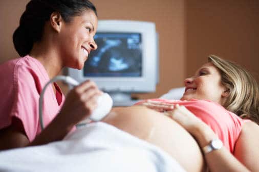 and to verify the expected date of delivery. It is usually done between 16 and 20 weeks of pregnancy.
For the procedure, a device that produces sound waves (transducer) is placed on the woman???s abdomen. The sound waves are processed to form an image that is displayed on a monitor. Sometimes, particularly during early pregnancy, the doctor uses an ultrasound device that can be inserted in the vagina. Ultrasonography produces high-quality images, including live-action images that show the fetus in motion. These images provide the doctor with useful information and can reassure a pregnant woman.
Ultrasonography can also be used to do the following:
- Show the fetus???s beating heart and thus confirm that the fetus is alive, as early as 6 weeks of pregnancy
- Identify the sex of the fetus, as early as 14 weeks of pregnancy
- See whether a woman is carrying more than one fetus
- Identify abnormalities, such as a mislocated placenta (placenta previa) or an abnormal position of the fetus
- Date the pregnancy and thus help determine whether the pregnancy is progressing normally
- Identify birth defects (sometimes)
- Guide the placement of instruments during certain procedures, such as prenatal diagnostic testing (see Prenatal Diagnostic Testing)
Toward the end of pregnancy, ultrasonography may be used to identify premature rupture of the fluid-filled membranes containing the fetus. Ultrasonography can provide information that helps doctors decide whether cesarean delivery is needed.
Immunizations
Experts recommend that all pregnant women be vaccinated against the influenza virus during the influenza (flu) season.
Pregnant women can be given the hepatitis B vaccine if needed.
Experts recommend a booster shot for tetanus, diphtheria, and pertussis (Tdap) after 20 weeks of pregnancy (preferably at 27 to 36 weeks) or after delivery, even if the shots are up-to-date.
Vaccines for measles, mumps, rubella, and varicella should not be given during pregnancy